Lowe syndrome

What is Lowe syndrome

Lowe syndrome is a hereditary disease characterized by three groups of symptoms: 1) eye manifestations, 2) central nervous manifestations, and 3) kidney manifestations. The disease is said to mostly occur in boys and very rarely in girls.


Diagnostic criteria

Symptoms of Lowe syndrome

Patients with Lowe’s syndrome present with three groups of symptoms listed below:
1)Eye manifestations
Abnormalities such as congenital cataract are observed. Congenital cataract is present bilaterally from the time of birth in almost all patients. The majority of patients are reported to have a vision of 0.2 or less.
2)Central nervous manifestations
Cognitive impairment, psychomotor retardation, convulsion, behavioral abnormalities, hypotonia, etc. are present.
There are differences among individuals in the degree of cognitive impairment and developmental retardation, but most commonly, the severity of the disorder is mild or moderate. It is said that many patients become able to walk independently at the age of 6 to 13 years. Convulsive seizure occurs in a little less than half of patients, but there are individual differences in the pattern of seizures. Behavioral abnormalities such as self-harm behavior are reported to occur as aging in about half of all patients.
3) Kidney manifestations
The dysfunction of a part of the kidneys called the proximal renal tubule causes the condition where amino acids, glucose, and electrolytes that cannot be reabsorbed leak into the urine (Fanconi syndrome). Consequently, symptoms such as failure to thrive, rickets, metabolic acidosis (the blood becomes acidic) may appear (see table below). In addition, the excretion of calcium into urine increases, and as a result, nephrocalcinosis is observed in about half of all patients. According to an investigation in Japan, patients develop end-stage kidney disease most commonly in their 30s to 40s.

Table Symptoms of Lowe syndrome based on a questionnaire survey in Japan
(Cited from “A Nationwide Survey on Hereditary Diseases Presenting with Tubular Proteinuria. ” Health and Labor Sciences Research Grants for FY 2015 to 2016 [Research Program on Rare and Intractable Diseases])


β2MG: a type of small molecular weight protein called β2microglobulin

Treatment for Lowe syndrome

Treatment is mainly symptomatic. Cataract often requires surgery. Convulsion is treated with anticonvulsant drugs, and muscle weakness is treated with physiotherapy. Various symptomatic treatments are given for kidney failure. There have also been some cases that progressed to end-stage kidney disease and required dialysis or kidney transplantation.

Q&A

1) What is the approximate number of patients with this disease?

It is estimated that the disease occurs in a few out of 100,000 boys, but the accurate frequency and number of patients are unknown.

2) Is the cause of this disease known?

OCRL on chromosome X has been identified as the responsible gene causing this syndrome.

3) Is this disease hereditary?

It is known to be an X chromosome recessive hereditary disease.
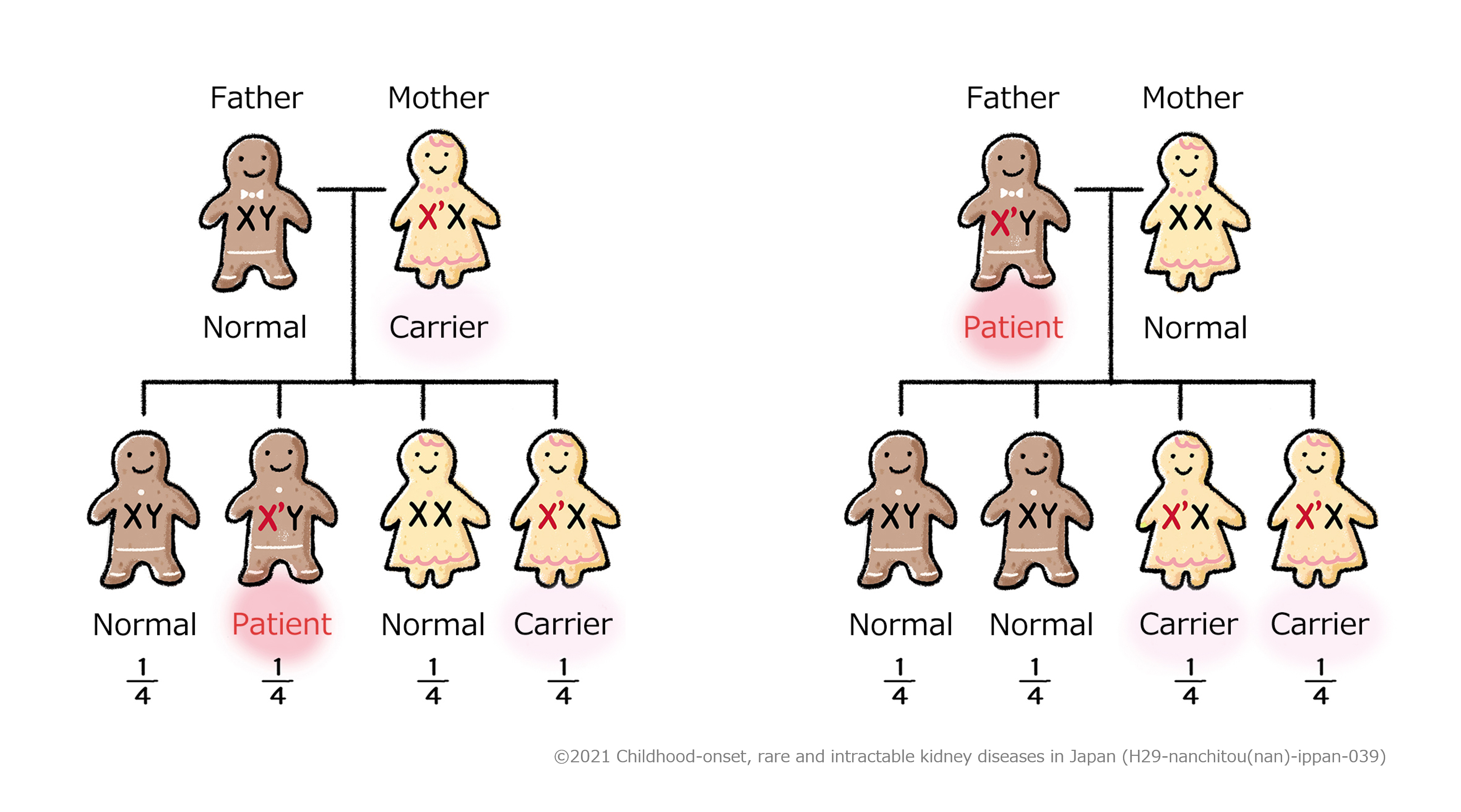
Explanation 
Being a carrier means that a person carries a gene that can cause a disease (X’ chromosome in the figure above) but does not develop the disease
In Figure 1, the mother carries an X’ chromosome causing the disease, but the other chromosome (X chromosome) is normal, and so she is a carrier but does not develop the disease. Among her children, if a son, who has only one X chromosome, inherits the X’ chromosome from her mother, he develops the disease. If a daughter inherits the X’ chromosome from her mother, the other chromosome is normal, so she becomes a carrier like her mother.
In Figure 2, the father has an X’ chromosome causing the disease, and this is the only X chromosome he has, and so he develops the disease. Among his children, because sons inherit a normal X chromosome from their mother, they are normal. On the other hand, girls inherit the X’ chromosome causing the disease from their father, and so they both become carriers.

4) What is the clinical course of this disease?

Treatment is mainly symptomatic. Renal dysfunction is progressive, and patients are reported to progress to end-stage kidney disease most commonly in their 30s to 40s.

Diagnosis and treatment guidelines
(For healthcare providers)

○ Overview

  • Overview
    Lowe syndrome (OMIM 309000), also known as oculo-cerebro-renal syndrome of Lowe (OCRL), is a disease characterized by three groups of symptoms: congenital bilateral cataract (glaucoma), central nervous manifestations, and kidney manifestations. It is caused by abnormalities in the OCRL gene encoding a phosphatidylinositol 4, 5-bis-phosphate-5-phosphatase (PtdIns(4, 5)P2). Lowe syndrome is an X chromosome recessive hereditary disease, and therefore, generally occurs in boys, but there have also been mild cases reported in girls caused by balanced X-autosome translocation. It is a very rare disease, with the frequency estimated to be approximately 1 in 500,000 to 1 million.
  • Causes
    OCRL (MIM 300535), which consists of 24 exons and is located on the long arm of the X chromosome, Xq25-26.1, has been identified as the responsible gene. OCRL protein encoded byOCRL, phosphatidylinositol 4, 5-bis-phosphate-5-phosphatase (PtdIns(4, 5)P2), is considered to act as a bioactive molecule in vivo and be involved in regulating various cell functions such as membrane transport, cell apoptosis, and cytoskeletal regulation. The abnormalities of the protein affect cytoskeletal remodeling and endocytosis, and this is thought to be the main pathological condition. However, the reason why target organs affected with clinical problems are limited though gene expression is observed generally in various organs and the detailed relationships between enzyme activity deficiency and various clinical symptoms are still unknown. The OCRL gene abnormalities cause not only Lowe syndrome, but also Dent disease (Dent disease-2). In Dent disease, low molecular weight proteinuria, hypercalciuria, nephrocalcinosis, etc. are observed, but cataract and mental retardation are absent. The detailed mechanism by which abnormalities in the same gene show different clinical features has not been found yet.
  • Symptoms
    The symptoms of Lowe syndrome are categorized into 3 major domains: eye manifestations, central nervous manifestations, and kidney manifestations.
    1)Eye manifestations
    Congenital cataract is present bilaterally from the time of birth in almost all patients. It is caused by migration defects in the lens epithelium, and since it occurs during the early stage of embryogenesis, it may be identified by fetal ultrasound scan. In addition, severe glaucoma accompanied by blepharitis and requiring surgical treatment usually occurs by 1 year of age in approximately 50% of child patients. Corneal scar and keloid without history of trauma usually occur in approximately 25% of patients under 5 years old. Worsened visual acuity due to partial retina degeneration occurs, and the majority of patients have a vision of 0.2 or less. Besides, corneal degeneration, nystagmus, strabismus, microphthalmos, etc. may be present. Female carriers are usually asymptomatic, but it is reported that fine cloudiness of the crystalline lens is observed by slit lamp microscope in at least 90% of patients.
    2)Central nervous manifestations
    All children have severe hypotonia with decreased deep tendon reflex at birth. Gross motor development is delayed, and 75% of patients become able to walk independently at the age of 6 to 13 years. The severity of mental retardation is moderate to severe with an IQ of 50 or less. Convulsive seizure occurs in 30 to 50% of patients, but there is no specific seizure pattern. Furthermore, behavioral abnormalities (self-harm behavior, irritability, autism, etc.) are reported to occur and worsen with aging. The electromyography is normal, and muscle biopsy reveals myopathy and neurogenic muscular atrophy. MRI often shows mild ventricular enlargement and periventricular cystic lesions.
    3)Kidney manifestations
    Kidney manifestations start with Fanconi syndrome occurring from the neonatal period to early infancy. Low molecular weight proteinuria and moderate albuminuria are always present, but urinary glucose is rarely present. Other kidney manifestations include aminoaciduria, proximal renal tubular acidosis due to failure of HCO3 reabsorption, and loss of phosphate, but the severity differs greatly among individuals (Table 1). In addition, hypercalciuria is often present, and nephrocalcinosis occurs in approximately 50% of patients. Regarding kidney function, the results of an international, retrospective cohort study were recently reported and showed that in 106 patients with abnormalities in the OCRL gene (88 patients with Lowe syndrome, 18 patients with Dent disease-2), decreased kidney function progressed faster in Lowe syndrome than in Dent disease-2. Nephrocalcinosis, hypercalciuria, and low molecular weight proteinuria were not interrelated with decreased kidney function.1)
    4) Other
    Hypophosphatemia/metabolic acidosis due to proximal renal tubular acidosis and hypotonia cause rickets/osteomalacia. In addition, body size at birth is within the normal range, but the final height is lower. Patients present with characteristic facial morphology such as prominent forehead and orbital hollow. Joint lesions (joint swelling, arthritis) and fibroma, dentigerous cyst, double dental arch, cryptorchidism, chronic constipation, scoliosis, and bleeding tendency may also be observed.
  • Diagnostic criteria
    The diagnosis of Lowe syndrome is suspected by three characteristic symptoms (congenital bilateral cataract, hypotonia, and Fanconi syndrome) and confirmed by the identification of the mutation in the OCRL gene.
    In a nationwide survey conducted in FY 2015 to 2016, 67 patients with Lowe’s syndrome were collected. Low molecular weight proteinuria (urine β2MG >5000 µg/L), cataract, and mental retardation were observed in 100% of patients (Table 1),2)and based on these results, the diagnostic criteria for Lowe syndrome were created.
  • Diagnostic criteria for Lowe syndrome

    A Symptoms
    1.Congenital cataract
    2.Central nervous manifestations (psychomotor retardation)

    B Laboratory findings
    1.Urineβ2MG >5000 µg/L

    C Differential diagnoses
    Dent disease, mitochondrial disorders, galactosemia, hereditary fructose intolerance, Fanconi-Bickel syndrome

    D Genetic testing
    1.Mutation in the OCRL gene

    <Diagnostic category>

    Definite:2 items of A + 1 item of B are met, differential diagnoses of C are excluded, and D is met.

    Probable:2 items of A + 1 item of B are met, and differential diagnoses of C are excluded.

  • Treatment
    1) Eye manifestations
    For cataract, lens extraction should be performed as early as possible. Taking into the account the possibility of concomitant glaucoma, artificial lenses are rarely used. Besides these, it is essential to measure intraocular pressure regularly.
    2) Central nervous manifestations
    For psychomotor retardation, appropriate and sufficient rehabilitation (development support) is necessary. After detailed evaluation, rehabilitation is provided at a local development support center or rehabilitation facility. Behavioral abnormalities (e.g., abnormal excitement, stereotypical behavior) may be present, and if they affect everyday life, drug therapy will be considered. In literature, atypical antipsychotic drugs (e.g., clomipramine, paroxetine, risperidone) are reported to be effective, and in Japan, risperidone and aripiprazole are easy to use among those covered by insurance. If the patient has epilepsy, treatment with an antiepileptic drug is required. When providing treatment with these drugs, drugs should be selected in consultation with a nephrologist by paying attention to kidney function. As for antiepileptic drugs, blood drug concentration can be measured, and efforts should be made to maintain effective concentration.
    3)Kidney manifestations
    The correction of proximal renal tubular acidosis is most important, and treatment with citric acid Na/K-salt or sodium bicarbonate is essential. Patients with rickets due to hypophosphatemia require treatment with active vitamin D and neutral phosphate. However, caution is needed for nephrocalcinosis due to overdose, and treatment should be given while monitoring the urine Ca/Cr ratio.
  • Prognosis
    Life expectancy is generally estimated to be in the 40s, and the leading causes of death are said to be progressive kidney failure and related complications. However, better understanding is needed about this because adult kidney function data are limited.
    Furthermore, there is no unified view on dialysis initiation or kidney transplantation. These interventions are sometimes not provided due to adherence issues or the family’s request, but there are reported rare cases of transplantation. Further surveys are needed on the improvement in vital prognosis by medical intervention

Table 1 Frequency of the clinical symptoms of Lowe syndrome based on a questionnaire survey in Japan
(Cited from “A Nationwide Survey on Hereditary Diseases Presenting with Tubular Proteinuria.” Health and Labor Sciences Research Grants for FY 2015 to 2016 [Research Program on Rare and Intractable Diseases])

References

1) Zaniew M, Bökenkamp A, Kolbuc M. et al.: Long-term renal outcome in children with OCRL mutations: retrospective analysis of a large international cohort. Nephrol Dial Transplant 33: 85-94. 2018
2) Miura K: A Nationwide Survey on Hereditary Diseases Presenting with Tubular Proteinuria. Health and Labor Sciences Research Grants for Research Program on Rare and Intractable Diseases, Annual Report FY 2016, 2016.

この病気に関する資料・関連リンク

ページトップへ